29 Sep Canine Acanthomatous Ameloblastoma (CAA) in the Dog
First things first: When you see an oral mass in the mouth of a dog or cat, biopsy it as soon as possible. Get a representative biopsy of the mass without interrupting the architecture around the mass. Smaller masses may be able to be removed completely, but PLEASE take clinical images of this in case your report comes back unfavorable. Telling a client that you’re going to ‘keep an eye on it’ is not good medicine and can even have fatal consequences.
Second: Use a reputable pathologist. Nothing against the state laboratory pathologist, but get a knowledgable oral pathologist to read your samples. I use CCOMP (Center for Comprehensive Oral and Maxillofacial Pathology) at the University of Wisconsin.
What Is Canine Acanthomatous Ameloblastoma?
Canine Acanthomatous Ameloblastoma (CAA) is an aggressive, benign epithelial odontogenic tumor. Not only does it affect the gingiva, but also the underlying and surrounding bone. These tumors do not metastasize, but they are very aggressive locally. There are studies to indicate that these tumors may rarely convert to squamous cell carcinomas after radiation therapy. This is a rare phenomenon.
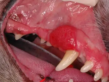
Right maxillary canine tooth with acanthomatous ameloblastoma
CAA Treatment Options
Treatment options are complete surgical removal of the mass, radiation therapy, and Bleomycin injection therapy. Surgery is still the gold standard of care and is extremely favorable providing you achieve clean surgical margins. Rostral mandibulectomy to spare the symphysis can be achieved with most cases and if a CAA is on the rostral mandible. A mandibular rim resection can be performed, sparing the ventral cortex with some CAA that involve the caudal mandible. With some cases, CT allows me to visualize the amount of bone destruction that conventional radiography may miss, so I always discuss this with pet owners.
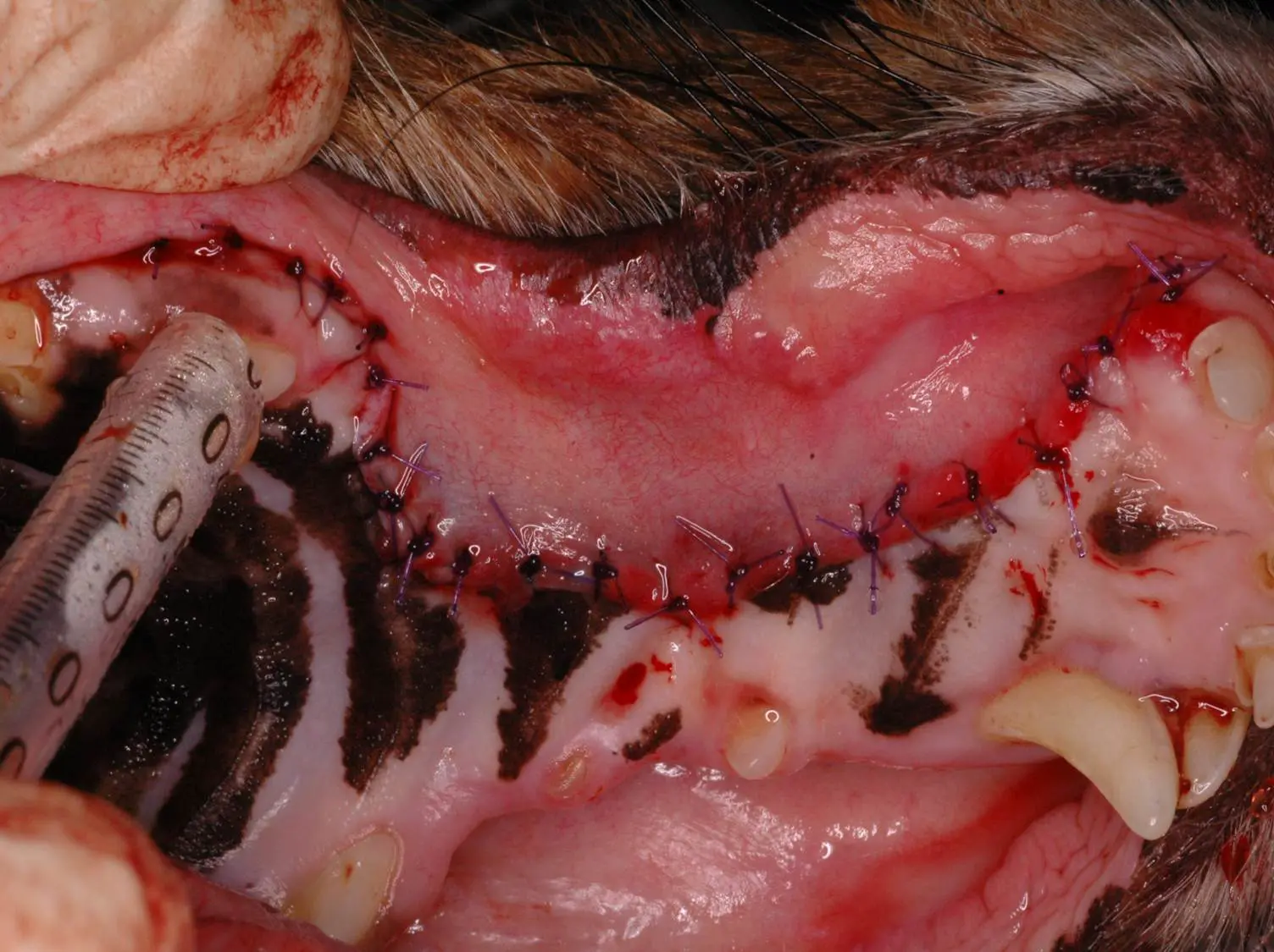
After surgical resection of above CAA on the right maxillary canine tooth. Margins were clean and the pet lived an uneventful life afterwards.
Thankfully, the outcome for this tumor is extremely favorable, providing it is diagnosed early and treatment performed at that time. But DO NOT try to debulk this mass on presentation. Remember to get an incisional biopsy and send it to a reputable pathologist.
Barden Greenfield, DVM, DAVDC – Your Pet Dentist of Nashville
(For those reading my constant contact blog, the answer is D!)
Images used under creative commons license – commercial use (11/21/2023). Photo by Justin Veenema on Unsplash