
13 Oct Do’s and Don’t’s With Regard to Oral Biopsies in the Dog and Cat
Oral tumors compose approximately 5-8% of all tumors in the dog and cat. That’s a considerable number and the most difficult aspect of this large percentage is that most people do not spend time looking in their pet’s mouth, so these masses grow unnoticed.
Understanding Oral Tumors in Pets
Many studies regarding dog and cat oral tumors show that size indeed matters with regard to removal. Surgical removal of growths less than 1.0 cm in diameter has a higher probability of clean margins (tumor-free at the margin) than those greater than 1.0 cm in diameter. This is especially true for those growths in the rostral (front) of the mouth, regardless of whether it is the mandible or maxilla.
Benign vs. Malignant Oral Tumors
Oral tumors can be benign or malignant. Just because an oral tumor is benign does not mean that it may not be clinically aggressive. Acanthomatous ameloblastoma, a benign odontogenic tumor, is extremely aggressive and left untreated, can cause significant maxillary or mandibular destruction. A malignant tumor may or may not invade bone, depending on the type of tumor. However, a malignant tumor such as a malignant melanoma, can spread to regional lymph nodes as well as to the lungs early in the tumor growth process. Therefore, early detection and treatment are critical with regard to life expectancy post-surgery +/- additional therapy (radiation therapy, chemotherapy).
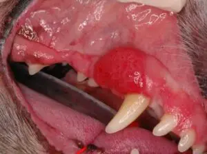
Acanthomatous ameloblastoma in the dog
The most common benign oral tumors are as follows:
- Acanthomatous ameloblastoma
- Periopheral odontogenic fibroma (POF)
- Periopheral odontogenic fibroma with ossification (POF/O)
The most common malignant oral tumors in dogs are as follows:
- Malignant melanoma
- Squamous cell carcinoma
- Fibrosarcoma
- Osteosarcoma
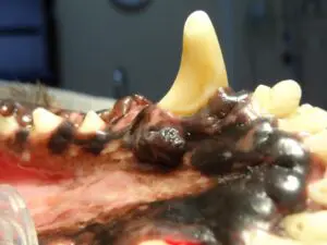
Oral melanoma in the dog
The most common malignant oral tumors in cats are as follows:
- Squamous cell carcinoma
- Fibrosarcoma
Do’s and Don’t’s of Diagnosing Dog and Cat Oral Tumors
Now let’s address the meat of the discussion, and that is how should one proceed when one sees an oral tumor in the mouth. This is extremely critical to understand oral surgical principles and what should be done and what should not be done. The first and foremost take home message is this: DO a biopsy first and submit it to a reputable pathologist.
Therefore, DO NOT try to remove the mass as it is without understanding what type of tumor it is. You will do tremendous harm to the pet if you try to remove it first. The reason is some tumors require a significant amount of margins to be cut (1.0 cm or greater) and this includes bone and underlying soft tissue. Once you cut a tumor without margins, it makes it extremely difficult for a veterinary dental specialist™ to know where the original tumor was located.
A critical step in this process once an oral tumor is identified is to inform the owner that a biopsy would be needed PRIOR to the removal of the mass. Once that mass has been identified by a pathologist, consultation with us is important. Most require advanced imaging such as Cone Beam CT (CBCT) to truly assess how much bone involvement is present. The Your Pet Dentist team has a CBCT machine and we routinely use this valuable tool on all our dental cases!
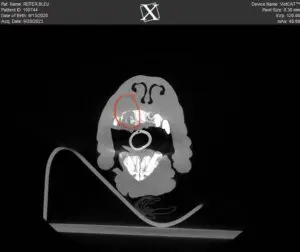
Cone Beam CT of an oral mass of the maxilla. Notice the red circular area indicating bone destruction of this mass. This allowed us to map out the margins necessary to achieve a clean surgical cut
A Step-by-Step Guide on Diagnosing Pet Oral Tumors
Below is the best step-by-step process needed once an oral tumor is identified:
- Take clinical pictures of the oral tumor and SAVE them in the pet’s medical record
- Perform dental radiographs (not survey images as they are non-diagnostic with regard to oral pathology)
- Take a biopsy of the oral tumor to get a true representative of the mass
- Submit to a reputable pathologist
- Once the biopsy returns from the pathologist, map out a plan going forward with the advice of a Veterinary Dental Specialist™
- A specialist can give you prognostic information, whether this mass spreads to other organs, etc…
DO NOT allow yourself to think you can cut without knowing what it is, and this is NOT a time to cut corners. Let’s put this into something a person can understand on the cellular level: A woman has felt a lump on her breast at home or picked up on a routine mammogram by her physician. The physician then recommends a radical mastectomy without biopsy. This woman will rightfully say NO, and find someone who can identify what the tumor is, how invasive it is, and then what options she has in front of her. That is the logical and wisest sequence one should follow. This same logical sequence should apply to oral tumors in your pet’s mouth. You will need to pay twice for anesthesia, but the consequences of a single cut and biopsy are loaded with danger looming. Don’t cut corners and make sure you follow the right sequence of events needed to provide the most adequate diagnosis for a pet.
Barden Greenfield, DVM, DAVDC Your Pet Dentist of Nashville
Images used under creative commons license – commercial use (10/13/2023). Photo by Helena Jankovičová Kováčová on Pexels