29 Dec What is the difference between Cone Beam CT and Dental Radiographs and Why does my pet need both?
The gold standard for dental imaging has long been digital intra-oral radiographs. This diagnostic tool gives us a 2-dimensional view of the hard tissues under the gumline. This includes the bone the tooth roots and other anatomic structures such as sinuses, foramina (where nerves and vessels enter/exit bone, and the temporo-mandibular joints (TMJ). Dental radiographs were first utilized in the 1950s. They allowed for dentists to truly assess tooth roots for changes associated with infection, inflammation, resorption/breakdown, and even fractures. They are vital to diagnostics, not only for our own dental health, but our pets as well. For many years, dental radiographs were the only modality that allowed us to be able to assess for tooth root abscesses, non-vital teeth, and any changes with the bone that support the teeth. Today, we’ll identify the difference between Cone Beam CT and Dental Radiographs and how they are used in veterinary dentistry.

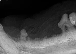
The image above shows one year old Deutsch Draahthaar with a supernumerary (extra) right mandibular 1st premolar that was unerupted and under the gumline. Impossible to know it existed without intra-oral imaging.
The Importance of Dental Radiographs
Two studies in the 90’s evaluated the additional findings that dental radiology allowed for in both cats and dogs. This study by Dr. Frank Verstrate confirmed what so many already knew, dental radiographs are not only useful but necessary and vital. They have become the gold standard for diagnosing oral and dental disease.
Advanced periodontal disease (stage 4 – >50% bone loss) and multiple retained tooth roots
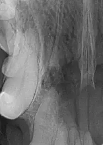
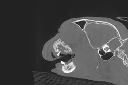
Large cystic lesion at rostral maxilla and incisors
CT Scans
Conventional CT or “CAT scans” have been utilized in the medical field since 1971. CTs allow for evaluation of anatomic structures in two different planes, transverse and sagittal. Conventional CT is extremely useful for soft tissue diseases, such as oral masses and other body cavities. However, the pixels and slice thickness does not allow for images detailed enough for most dental disease. Cone beam CT was first used in the late 1990s. This modality utilizes voxels which evaluate anatomic structures in 3 different planes, allowing for a full 3D evaluation of anatomic structures. This, due to the utilization of voxels and not pixels, along with a much smaller slice thickness, makes the cone beam CT ideal for evaluating dental structures and hard tissues of the head/neck.
Numerous studies have proven the advanced diagnostic yield of cone beam CT compared to intra-oral dental radiographs. In fact, cone beam CT is capable of allowing for the diagnosis of a tooth root abscess (periapical lesion) in as little as 14 days compared to 60 days for intra-oral radiographs. Not to mention, evaluating 3D structures such as tooth roots, bone, and other hard structures associated with the oral cavity, in a 3D image allows for a new degree of accuracy. Cone beam CT technology also gives the user the ability to reconstruct the image into a full 3-dimensional image. This allows for true evaluation of hard tissues and possible changes or defects such as those found with oral masses. Cone beam CT is quickly becoming the gold standard for veterinary dentists and oral surgeons.

Left – right caudal maxilla in an 8 year old cat. Right – same cat with CBCT imaging. Significant more bony changes are evident on the CT image
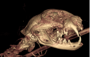
3D reconstruction of a cat with an oral tumor causing severe bone loss
The Importance of Utilizing Radiographs & CT Scans
So Cone beam CT sounds perfect, right!? Of course that’s all the imaging your pet would need for diagnostics, right? Unfortunately that isn’t quite right. Cone beam CT absolutely has its limitations. Its greatest limitation is the inability to evaluate soft tissue structures as well as it does hard tissue structures. What this means is, cone beam CTs are not ideal soft tissue masses, evaluating muscle around the hard tissues, or even salivary glands within the head and neck. Additionally, cone beam CT may affect the evaluator’s ability to accurately diagnose non-vital teeth strictly on pulp cavity width along with specific types of tooth resorption. There are often cases where both intra-oral radiographs and cone beam CT are necessary to fully diagnose a lesion or oral disease. As software associated with cone beam CT advances, this may not always be the case.
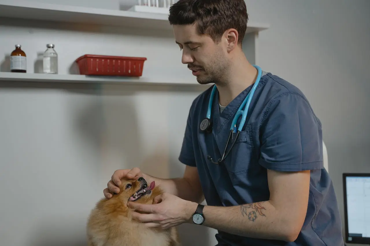
State-of-the-Art Veterinary Dentistry Clinic in Nashville
The team at Your Pet Dentist was the first private practice facility in the state of Tennessee to have both Cone Beam CT and full intra-oral radiography capabilities (with a different practice in Knoxville offering the same imaging options). Drs Briggs and Greenfield utilize these modalities on every patient that undergoes an oral or dental procedure. They enjoying not only the benefits these imaging modalities allow them to see and evaluate, but also enjoy showing you and your family these often cool and surprising images. If it has been recommended that your pet undergo an anesthetic dental procedure, intra-oral imaging is not only key but truly a necessity to evaluate the teeth and oral structures. Do not hesitate to ask your regular veterinarian or our team at Your Pet Dentist more about these imaging modalities!
Photo by Tima Miroschnichenko from Pexels